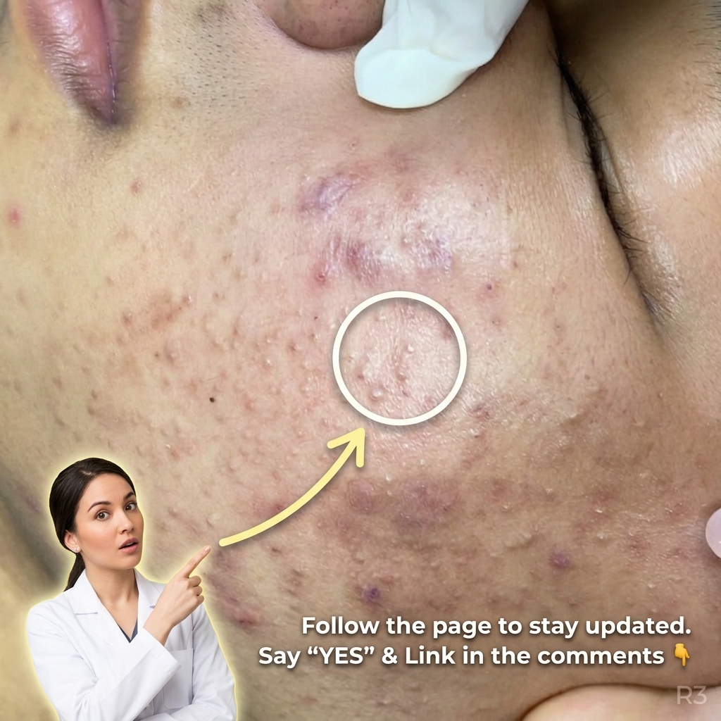
The development of acne begins deep within the pilosebaceous unit, where an overproduction of sebum combines with abnormal keratinization. When dead skin cells fail to shed properly, they accumulate inside the follicular duct, leading to clogged pores. This trapped mixture creates an ideal anaerobic environment for follicular microflora, particularly Cutibacterium acnes, to proliferate. In response to bacterial overgrowth and accumulated lipids, the innate immune system triggers a localized inflammatory cascade. Early non-inflammatory lesions, such as open comedones known as blackheads and closed comedones known as whiteheads, can quickly evolve into inflamed papules and pustules as immune cells infiltrate the area. Oily skin often exacerbates this process by providing a continuous supply of substrates that maintain follicular congestion. Understanding this initial follicular disruption is essential because the intensity and duration of the underlying dermal inflammation directly dictate the subsequent structural and pigmentary changes observed in the surrounding tissue.
Post-inflammatory hyperpigmentation arises as a direct biological consequence of the skin's inflammatory response to acne lesions. When an acne papule or cyst breaks down the follicular wall, inflammatory mediators including prostaglandins, leukotrienes, and interleukins are released into the surrounding epidermal and dermal layers. These signaling molecules act as potent stimulants on epidermal melanocytes, the specialized pigment-producing cells located along the basal layer of the epidermis. In response to inflammatory stress, melanocytes undergo upregulated melanogenesis, producing excess melanin pigment. This synthesized melanin is subsequently transferred to neighboring keratinocytes or, if the basal membrane is disrupted, leaks into the upper dermis where it is engulfed by dermal macrophages. The resulting dark spots represent stored cellular pigment left behind long after the active acne lesion, such as an inflamed whitehead or cyst, has resolved.
Several endogenous and exogenous factors influence the severity and persistence of post-inflammatory hyperpigmentation following an acne flare. Individual skin phototypes play a significant role, as individuals with higher melanin baselines often exhibit more reactive melanocytes upon inflammatory stimulation. Furthermore, physical manipulation of acne lesions—such as squeezing, picking, or aggressive extraction of blackheads and clogged pores—exacerbates mechanical trauma to the follicular wall. This extra trauma deepens the inflammatory cascade, driving pigment deeper into the dermal layers and extending the resolution timeline. Ultraviolet radiation from sun exposure serves as another critical aggravating factor; UV light directly stimulates melanocytes to continue producing pigment, turning faint marks into persistent discoloration. Additionally, an impaired skin barrier can heighten cutaneous sensitivity, prolonging local inflammatory responses and rendering the tissue more susceptible to lingering hyperpigmentation.
Addressing both active acne and post-inflammatory hyperpigmentation requires topical strategies that simultaneously target follicular occlusion and melanin production pathways. Ingredients like topical retinoids are frequently discussed in dermatological literature because they promote cellular turnover and normalize keratinization, helping to clear clogged pores while gradually dispersing accumulated pigment in upper epidermal layers. Chemical exfoliants, such as lipophilic salicylic acid, effectively penetrate oily skin to clear debris from within the pores, mitigating the inflammatory triggers that precede hyperpigmentation. Additionally, targeted brighteners like azelaic acid and niacinamide offer dual benefits: azelaic acid functions as a mild antibacterial agent and a tyrosinase inhibitor, curbing abnormal melanin synthesis, while niacinamide helps suppress melanosome transfer from melanocytes to keratinocytes. Incorporating broad-spectrum sun protection remains an essential complementary step, preventing UV rays from darkening resolving inflammatory marks.
Long-term care for acne-prone skin prone to hyperpigmentation centers on maintaining a delicate balance between active treatment and skin barrier preservation. Over-treating active lesions with harsh physical scrubs or excessive drying agents can cause micro-irritation, which paradoxically triggers secondary inflammation and worsens pigment deposition. Maintaining a healthy skin barrier and balanced cutaneous microbiome helps protect against external pathogens while supporting natural epidermal repair mechanisms. Consistency is particularly vital, as cellular turnover and dermal pigment clearance occur gradually over several weeks to months. A thoughtful, gentle skincare routine focused on sebum regulation, controlled exfoliation, and rigorous sun protection allows the tissue to recover without invoking unnecessary cellular stress. When hyperpigmentation remains resistant to non-prescription approaches, evaluation by a qualified dermatologist can help clarify individual needs and provide targeted clinical guidance.
Related acne topics
- Clinical Approaches to Distinguishing and Managing Comedonal and Inflammatory Acne
- Understanding the Impact of Occlusive Skincare Products on Pore Congestion and Long-Term Skin Dynamics
Health information notice: This article is for general informational and educational purposes only. It is not medical advice and does not replace consultation with a licensed dermatologist or healthcare provider.
This article was generated with AI assistance and has not been individually reviewed by a medical professional. View the accompanying YouTube video.
